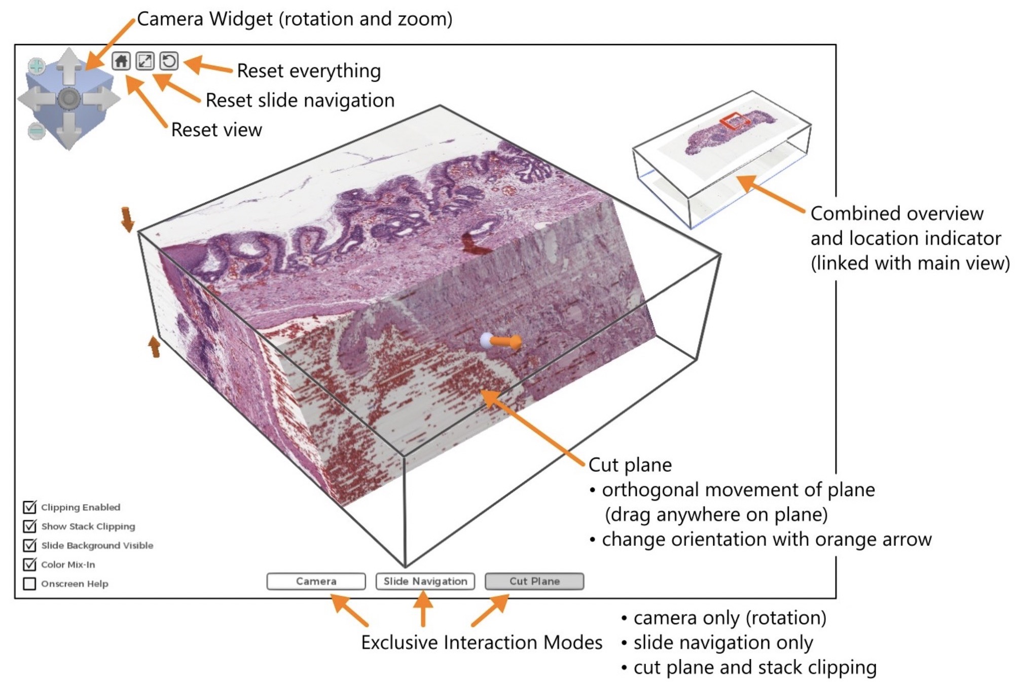
Histology, the medical analysis at microscopic level of tissue specimen from the human body, is a cornerstone in medical research. Digitization of the microscopy images opens up exciting possibilities for visualization and image analysis. A particularly exciting area for research is correlating the structure of 3D histology with in vivo imaging findings of the same tissue (before surgery/biopsy), such as Computed Tomography (CT) or Magnetic Resonance (MR) imaging. A system providing novel tools for visualization of 3D histology including effective volume rendering and interactive feature exploration methods has been developed. A user study with positive feedback has been performed with participants from two labs in Sweden and UK. A paper describing the system and the study results has been published in the most prominent conference and journal in the visualization domain. The software is implemented in the Inviwo framework and is available for research groups in the area.
Reference: Interactive Visualization of 3D Histopathology in Native Resolution. M. Falk, A. Ynnerman, D. Treanor, C. Lundström. IEEE transactions on visualization and computer graphics, 2018.


